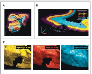
Characterizing the precise pathology in autistic spectrum disorders, let alone other psychiatric disorders, has remained challenging. Postmortem studies of actual brain tissue are rare but have the potential to provide important clues regarding specific abnormalities in neurodevelopment that are not detectable through other means such as neuroimaging. This major study published in the New England Journal of Medicine provides direct and provocative evidence of specific neuronal pathology that may underlie some of the core features of autism. 
The authors used RNA in situ hybridization techniques in order to look for gene expression patterns of 25 potential genes that have been implicated in the pathogenesis of autism and mark the presence of particular types of cells at different layers in the cortex. These analyses were performed on areas within the frontal, temporal, and occipital lobes of 11 children with autism and 11 children controls from two established brain banks. Children were between 2 and 15 years of age and the autistic sample was predominantly male.
In 10 out of 11 children with autism, compared to 1 out of 11 children without it, the study found focal areas of significant cytoarchitectural disorganization in the prefrontal and temporal cortex but not in the occipital cortex. These areas were referred to as “patches” and tended to be between 5 and 7mm long, often existing right next to unaffected cortex. The dysfunction was found for different types of neurons but not glial cells. While there was variability found in terms of the precise types of cells and cortical layers that were most affected in the autistic group, such a relatively unique and consistent finding is a major step forward towards understand the precise brain differences in children with autistic spectrum disorder. The data also strongly points to autism beginning in the prenatal period when cortical layering and cellular differentiation are occurring.
The authors wrote that they were somewhat surprised to see such as similar pathological feature across so many children with autism given its heterogeneous nature. They also note that while the presence of these patches was evident, their study does not suggest a mechanism for how these abnormalities in neuronal migration and differentiation they develop.
For those interested in visualizing these brain changes, the NIMH released a YouTube video that shows what the patches look like. I would definitely recommend looking at this to better flavor of these intriguing findings.
Reference
Stoner R, et al. Patches of disorganization in the neocortex of children with autism. NEJM 2014;370:1209-1219.
Tags: autism, autistic spectrum disorder, brain

